Get More Information:
What is a Lung Nodule?
Minimally Invasive Diagnostic Procedures (Testing)
Radiation Therapy for Lung Cancer
Find Lung Cancer Clinical Trials
Lung Cancer Screening
Learn about our screening program
to promote early detection for those at high risk of developing lung cancer.
Listen In
The Cancer Institute’s Dr. John Langenfeld explains the benefits of lung cancer screening in this WOR radio interview. Click here to listen
Patients found to have a lung nodule, enlarged lymph nodes, or other abnormalities in their chest often require tissue to be obtained so a diagnosis can be determined. The Lung Cancer/Thoracic Oncology Program at Rutgers Cancer Institute has state-of-the-art minimally invasive diagnostic tools and therapeutic procedures needed to effectively diagnose and treat patients. Obtaining sufficient tissue is an important first step in determining the best treatment plan.
Experts in lung disease, our highly trained team of surgeons, pulmonologists, medical oncologists, radiation oncologists, nurses, and other personnel can help you select the diagnostic test that is best for you. Diagnostic tools available at the Cancer Institute include:
Fiber Optic Bronchoscopy
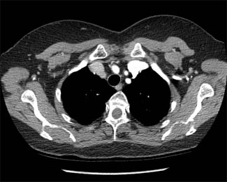
Bronchoscopies are used to look into the airways of patients and are performed to obtain cultures for pneumonias, evaluate for bleeding, and to biopsy a lung nodule or mass within the major airways. Bronchoscopes use fiber optic cables that allow magnification and viewing inside of the major airways. The major airways include the trachea (windpipe) and its branches leading into the lung. When airways enter into the lung tissue they become too small to be reached by a conventional fiber optic bronchoscope. Newer technology such as navigational bronchoscopy allows “visualization” of these smaller airways.
Navigational Bronchoscopy
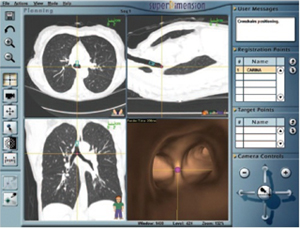 Navigational bronchoscopy is an exciting new technology that enhances our ability to make diagnosis of diseases in the chest. The technology applies an electromagnetic guidance system to a fiber optic bronchoscope together with enhanced computerized imaging to guide the physician directly to the abnormality within the lung. Navigational bronchoscopy allows doctors to biopsy abnormalities within the lung using a minimally invasive technique. The addition of confocal microscopy together with a navigational bronchoscopy allows direct visualization of the tissue increasing diagnostic yields. We are among a select group of centers in the state that use confocal microscopy. We are the only center in New Jersey using confocal microscopy. These minimally invasive technologies provide a great advantage to the patient and physician to obtain a diagnosis and determine a treatment plan.
Endobronchial Ultrasound (EBUS)
Endobronchial Ultrasound (EBUS) adds ultrasound to fiber optic bronchoscopes. Fiber optic bronchoscopes can only visualize the inside of the large major airways leading into the lung. At times enlarged lymph nodes or other masses surrounding the major airways need to be biopsied to determine the best treatment. Enlarged lymph nodes or masses surrounding the major airways cannot be seen with fiber optic bronchoscopes. The EBUS allows the physician to “visualize” outside the walls of the major airways allowing for an accurate biopsy. EBUS is an important tool for diagnosis, staging of lung cancer, and determining the most appropriate treatment for you.
Mediastinoscopy
Mediastinoscopy is a minimally invasive technique using fiber optic technology that can obtain larger amounts of enlarged lymph nodes or mass surrounding the major airways. Obtaining larger pieces of lymph nodes increases allows for further studies such as flow cytometry and genetic sequencing, which are sometimes needed. Mediastinoscopy is performed through a small 1.5 cm incision at the base of the neck under anesthesia. Mediastinoscopy is a same day procedure that is quick, safe, and has a fast recovery.
Video Assisted Thorascopic Surgery (VATS)
Lung nodules that are highly suspicious for cancer should be removed through a minimally invasive lung surgery called Video Assisted Thorascopic Surgery or VATS. Since the whole nodule is removed and examined it is the most definitive means to make a diagnosis of a lung nodule. VATS uses a video camera that is inserted into the chest through one of four small incisions. If only the lung nodule is removed patients typically are sent home the next day. This minimally invasive approach provides easy recovery with minimal discomfort.
Endobronchial Tumors
At times, tumors can grow into the major airways inhibiting the ability to breath. Removing the blocking tumor can significantly improve breathing. Doctors can remove the tumor using different lasers and cryotherapy. Treatment to prevent the recurrence of the tumor includes airway stents and the placement of local endoscopic radiation.